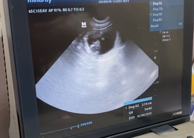
If your pet is under the weather and your vet has recommended an ultrasound, your first thought might be of pregnancy. While checking in on growing puppies or kittens is certainly one use, ultrasonography in veterinary medicine is actually a powerhouse diagnostic tool used to solve all kinds of internal mysteries.
Unlike X-rays, which show the general shape and placement of bones and solid organs, an ultrasound lets your vet look inside the organs in real-time. It is the difference between looking at a static photograph of a house (an X-ray) and walking through the front door to see what is happening in the living room (an ultrasound).
Let's dive into how this gentle, pain-free technology works, what it can find, and what to expect when your pet goes in for a scan.
How Does Veterinary Ultrasound Work?
Ultrasonography is completely non-invasive and uses no radiation. Instead, it relies on high-frequency sound waves to create a live picture.
During the procedure, a handheld probe called a transducer is pressed against your pet's skin. The transducer sends harmless sound waves into the body, which bounce off internal organs and tissues. The probe catches the returning "echoes," and a computer translates those echoes into a live, moving image on a screen.
Because different tissues (like fluid, muscle, and bone) bounce sound waves back differently, vets can see incredibly detailed structures—even down to the thickness of a cat's intestinal walls or the squeeze of a dog's beating heart.
X-Rays vs. Ultrasound: What's the Difference?
Vets often use these two tools together because they complement each other perfectly:
- X-Rays (Radiographs): Best for bones, joints, gas-filled lungs, and swallowed metallic objects. Provides static, 2D flat images.
- Ultrasound (Ultrasonography): Best for soft tissues, fluid-filled organs, and moving structures. Provides live, moving, multi-dimensional video.
Why Might Your Vet Recommend an Ultrasound?
Because it provides such detailed images of soft tissues, ultrasound is highly versatile. Vets commonly use it for:
1. Abdominal Scans
If your pet has chronic vomiting, diarrhea, unexplained weight loss, or abnormal blood work, an abdominal ultrasound is the gold standard. It allows the vet to examine:
- The Liver & Spleen: Looking for changes in size, texture, or the presence of tumors.
- The Kidneys & Bladder: Searching for bladder stones, structural changes, or signs of chronic kidney disease.
- The GI Tract: Measuring the thickness of the stomach and intestinal walls (critical for diagnosing Inflammatory Bowel Disease or lymphoma) and checking for blockages.
2. Echocardiograms (Heart Ultrasounds)
When a vet hears a heart murmur or suspects heart disease, they will perform an echocardiogram. This specialized scan lets them watch the heart valves open and close, measure the thickness of the heart walls, and track the flow of blood in real-time.
3. Safe Biopsies and Aspirates
If a vet spots a mass on an internal organ, they can use the ultrasound as a live guide. By watching the screen, they can safely guide a tiny needle directly to the mass to collect cells (a fine-needle aspirate) without needing to perform invasive exploratory surgery.
What to Expect: Preparing Your Pet for the Scan
Fortunately, an ultrasound is a highly gentle procedure. For most pets, it feels like getting a slightly cool belly massage! Here is what the process usually looks like:
- The Fasting Window: You will likely be asked to withhold food before the scan. A stomach full of digesting food and gas blocks sound waves, which can completely obscure the organs behind it. (Water is usually fine!)
- Getting Shaved: To get a clear picture, the transducer must make direct contact with the skin. Because fur traps air—the ultimate enemy of ultrasound waves—your pet's belly or chest will need to be shaved.
- Positioning & Gel: Your pet is gently placed in a padded, V-shaped cushion. A technician will hold and comfort them while a cool, water-soluble gel is applied to the shaved area to help transmit the sound waves.
- The Scan & Results: The vet or a veterinary radiologist glides the probe over the area, capturing images and measurements. Once finished, the gel is wiped off, and your pet is ready to go home—often with immediate answers for you!
Do They Need Sedation?
In 90% of cases, no anesthesia or sedation is required because the procedure is entirely painless. However, if your pet is extremely anxious, painful, or needs to lie perfectly still for a delicate needle biopsy, your vet may recommend a mild, safe sedative to keep them relaxed and comfortable.
Ultimately, ultrasonography is one of the safest and most detailed diagnostic tools available in modern veterinary care. It takes away the guesswork, allowing your veterinary team to target treatments precisely and get your furry friend back on the road to recovery as quickly as possible.