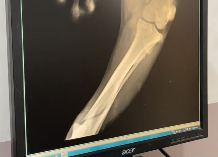
If your pet is limping, coughing, or has accidentally swallowed something they shouldn't have, your veterinarian's first recommendation will likely be to "take some rads." While "radiograph" is the formal medical term, most of us know them by their everyday name: X-rays.
As one of the oldest, most reliable, and fastest diagnostic tools in veterinary medicine, radiographs give your vet an immediate look at the structural layout of your pet's body. Let's explore how veterinary X-rays work, what they can diagnose, and what actually goes on behind the scenes when your pet goes into the radiology room.
How Do Veterinary Radiographs Work?
At its core, a radiograph is a shadow picture. X-rays are a form of high-energy electromagnetic radiation. When the X-ray machine is activated, a safe, highly controlled beam of radiation passes through your pet's body and hits a digital sensor plate underneath them.
Different tissues in your pet's body absorb different amounts of radiation:
- Bones (Dense tissue): Absorb almost all the radiation. Very little reaches the sensor plate, which is why bones show up as bright white.
- Air/Gas (Very low density): The radiation passes straight through to the sensor. This makes air-filled structures (like the lungs) show up as black.
- Soft Organs & Fluids (Medium density): Absorb some radiation, showing up in various shades of gray.
By reading these varying shades of white, black, and gray, your veterinarian can spot anomalies, structural damage, or fluid accumulation.
What Can Radiographs Diagnose?
Radiographs are the ultimate "first responder" of veterinary imaging. They are incredibly useful for diagnosing a wide array of conditions across different parts of the body:
1. Orthopedic Issues (Bones and Joints)
- Fractures and Breaks: Pinpointing the exact location and severity of broken bones.
- Arthritis and Joint Disease: Looking for bone spurs, joint swelling, or narrowing spaces between joints (like in hip dysplasia).
- Bone Tumors: Identifying areas where bone tissue is abnormally wearing away or growing too fast.
2. Chest Anomalies (Heart and Lungs)
- Pneumonia or Asthma: Spotting fluid, mucus, or inflammation in the lungs.
- Congestive Heart Failure: Checking if the heart silhouette is abnormally enlarged or if fluid is backing up into the lung cavity.
3. Abdominal Mysteries
- Foreign Body Ingestion: Locating swallowed coins, rocks, metal toys, or bones. (Soft items like socks, plastic wrap, or hairballs may require an ultrasound!)
- Organ Enlargement: Checking the overall size of the liver, spleen, or kidneys.
- Bladder Stones: High-density mineral stones in the bladder show up as bright white spots.
What Happens Behind the Closed Doors?
It is completely normal to wonder what happens when your pet is led into the X-ray room. Because we have to protect human workers from repetitive, daily radiation exposure, pet owners are generally not allowed inside the room during the scan. Here is the step-by-step process of what your pet experiences:
- Safety Gear for Staff: Safety first. The veterinary team wears lead-lined aprons, thyroid shields, and lead gloves to protect themselves from scattered radiation.
- Positioning the Patient: Your pet is gently lifted onto the metal X-ray table. The technicians place them in very specific positions (usually lying on their side or their back) to get the exact views the vet needs. Foam blocks, sandbags, or gentle tie-downs are often used to help keep them steady without holding them tightly.
- The Exposure: The staff step back, align the X-ray beam, and take the picture. Just like with a camera, the pet must be completely still for a split second, or the image will blur.
- Instant Analysis: Because almost all modern clinics use digital radiography, the image appears on the computer screen within seconds. The vet can zoom in, adjust the contrast, and make an immediate assessment.
FAQs: Common Parent Concerns
- Is the radiation dangerous for my pet? No. The dose of radiation used for a standard diagnostic pet X-ray is extremely low and completely safe. Because pets do not get X-rays daily, the risk of any adverse health effects from the radiation itself is virtually zero.
- Will my pet need to be sedated? Most calm pets can have X-rays taken with just some gentle, manual reassurance from the team. However, sedation is highly recommended if: Your pet is in severe pain (such as from a broken bone) and moving them into position hurts; Your pet is highly anxious, aggressive, or struggling, which makes taking a clear, unblurred image impossible; The vet needs to take precise joint measurements (like checking for hip dysplasia), which requires the muscles to be completely relaxed.
By giving your vet a clear, immediate look inside, radiographs take the guesswork out of treatment—ensuring your pet gets the precise care they need, right when they need it.